High-resolution 3T MR neurography of the brachial plexus and its branches, with emphasis on 3D imaging

125Citations
Citations of this article
176Readers
Mendeley users who have this article in their library.

Abstract

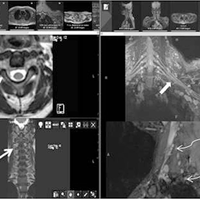
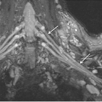
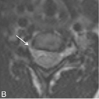
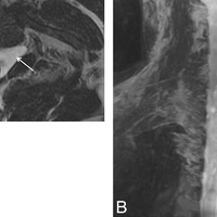
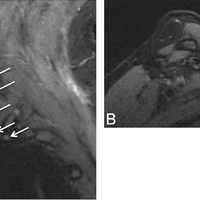
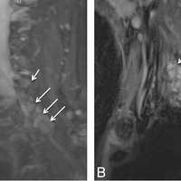
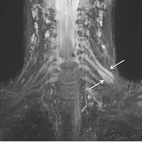
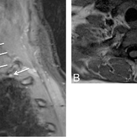
With advancement in 3D imaging, better fat-suppression techniques, and superior coil designs for MR imaging and the increasing availability and use of 3T magnets, the visualization of the complexity of the brachial plexus has become facile. The relevant imaging findings are described for normal and pathologic conditions of the brachial plexus. These radiologic findings are supported by clinical and/or EMG/surgical data, and corresponding high-resolution MR neurography images are illustrated. Because the brachial plexus can be affected by a plethora of pathologies, resulting in often serious and disabling complications, a better radiologic insight has great potential in aiding physicians in rendering superior services to patients.

Figures

  • FIG 1. MRN technique. 3T MR neurography imaging sequences with isotropic multiplanar reconstruction. 3D T2 SPACE with multiplanar reconstruction of the cervical spine (medium arrows ). Axial T1-weighted image shows the bilateral brachial plexus through the lower aspect of the scalene triangle (small arrows ). Coronal MIP 3D STIR SPACE image shows the brachial plexus (thick arrow ). Sequential sagittal STIR images show the normal and symmetric C5-T1 nerve roots (curved arrows ), trunks (circle ), and cords (oval ).
  • FIG 2. Normal brachial plexus. Coronal MIP 3D STIR SPACE image focused on the left side shows the anatomy of the brachial plexus (arrows).
  • FIG 3. IsolatedC6 radiculopathy. A 51-year-oldwomanwith right armpain and a tingling sensation, clinically suspected of having brachial plexitis versus radiculopathy. Sagittal STIR (A ), axial T2 SPACE (B ), and coronal 3D MIP STIR SPACE (C ) images show an asymmetrically hyperintense and diffusely enlarged isolatedC6 nerve root (arrows ), corresponding to themarkedly narrowed right C6 neural foramen. The findings are in keeping with cervical radiculopathy, in the setting of cervical spondylosis.
  • FIG 4. Nerve root avulsion. A 22-year-old man after a motor vehicle crash and ulnar-sided arm weakness. Axial T2-weighted (A ) and coronal MIP 3D STIR SPACE (B ) images show the avulsed left T1 nerve root (large arrows ) and C8 nerve root (small arrow ) with pseudomeningocele formation.
  • FIG 5. Stretch injury with suprascapular neuropathy. A 48-year-old man after a recent motor vehicle crash. Sequential sagittal STIR images (A and B ) show asymmetrically enlarged and hyperintense C5 and C6 nerve roots (large arrows in A ) relative to other normal nerve roots (small arrows ). All the nerve segments are in continuity. Notice the hyperintense suprascapular nerve (large arrow in B ) and mild denervation edema of the supraspinatus and infraspinatus muscles (stars ).
  • FIG 6. Multiple acute nerve root avulsions. A 51-year-old man with loss of function in the left upper extremity due to recent motor vehicle crash. Coronal MIP 3D STIR SPACE (A ) and sagittal STIR (B ) images show avulsed C6, C7, and C8 nerve roots (arrows ) with abnormal morphology due to hemorrhage and edema from a recent injury, obscuring clear details of the nerve roots. Notice the normal C5 nerve root (large arrows in A ) and the normal T1 nerve root (small arrow in B ).
  • FIG 7. Neurotmesis. A 43-year-oldwomanwith loss of function in the left upper extremity following recent neck surgery. Coronal MIP 3D STIR SPACE image shows severed, enlarged, and hyperintense C5 and C6 nerve roots with distal end bulb neuromas (arrows), just proximal to the formation of the left upper trunk. The findings were confirmed on re-exploration, and the patient underwent immediate nerve transfer.
  • FIG 8. TOS with C8 neuropathy. A 17-year-old girl (trumpet player) with left arm and hand weakness in an ulnar distribution, exacerbated by shoulder abduction. Sagittal STIR (A ) image shows an asymmetrically enlarged and hyperintenseC8 nerve root (large arrow ) comparedwith the other normal nerve roots (small arrows ). Notice minimal asymmetric hyperintensity of the T1 nerve root, a common nonspecific finding. Axial T1-weighted (B ) MR image through the lower neck shows a prominent flow void (curved arrow ) indenting the left C8 nerve root (large arrow ). The EMG findings were normal in this case. On surgery, the flow void turned out to be a prominent branch of the left thyrocervical trunk, which was ligated. C8 neurolysis was also performed. The patient’s symptoms completely resolved following surgery.

References Powered by Scopus

Register to see more suggestions

Mendeley helps you to discover research relevant for your work.

Already have an account?

Cite

CITATION STYLE

APA

Chhabra, A., Thawait, G. K., Soldatos, T., Thakkar, R. S., Del Grande, F., Chalian, M., & Carrino, J. A. (2013, March). High-resolution 3T MR neurography of the brachial plexus and its branches, with emphasis on 3D imaging. American Journal of Neuroradiology. https://doi.org/10.3174/ajnr.A3287

Readers over time

‘12‘13‘14‘15‘16‘17‘18‘19‘20‘21‘22‘23‘24‘2508162432

Readers' Seniority

Tooltip

PhD / Post grad / Masters / Doc 57

49%

Researcher 27

23%

Professor / Associate Prof. 26

22%

Lecturer / Post doc 7

6%

Readers' Discipline

Tooltip

Medicine and Dentistry 100

81%

Neuroscience 12

10%

Nursing and Health Professions 7

6%

Agricultural and Biological Sciences 5

4%

Article Metrics

Tooltip
Mentions
References: 1
Social Media
Shares, Likes & Comments: 25

Save time finding and organizing research with Mendeley

Sign up for free
0