Prion-seeding activity in cerebrospinal fluid of deer with chronic wasting disease

48Citations
Citations of this article
39Readers
Mendeley users who have this article in their library.

Abstract

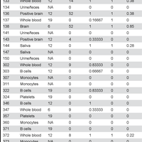
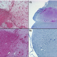
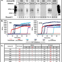
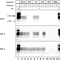
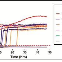
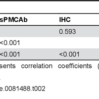
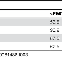
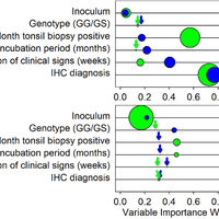
Transmissible spongiform encephalopathies (TSEs), or prion diseases, are a uniformly fatal family of neurodegenerative diseases in mammals that includes chronic wasting disease (CWD) of cervids. The early and ante-mortem identification of TSE-infected individuals using conventional western blotting or immunohistochemistry (IHC) has proven difficult, as the levels of infectious prions in readily obtainable samples, including blood and bodily fluids, are typically beyond the limits of detection. The development of amplification-based seeding assays has been instrumental in the detection of low levels of infectious prions in clinical samples. In the present study, we evaluated the cerebrospinal fluid (CSF) of CWD-exposed (n=44) and naïve (n=4) deer (n=48 total) for CWD prions (PrPd) using two amplification assays: serial protein misfolding cyclic amplification with polytetrafluoroethylene beads (sPMCAb) and real-time quaking induced conversion (RT-QuIC) employing a truncated Syrian hamster recombinant protein substrate. Samples were evaluated blindly in parallel with appropriate positive and negative controls. Results from amplification assays were compared to one another and to obex immunohistochemistry, and were correlated to available clinical histories including CWD inoculum source (e.g. saliva, blood), genotype, survival period, and duration of clinical signs. We found that both sPMCAb and RT-QuIC were capable of amplifying CWD prions from cervid CSF, and results correlated well with one another. Prion seeding activity in either assay was observed in approximately 50% of deer with PrPd detected by IHC in the obex region of the brain. Important predictors of amplification included duration of clinical signs and time of first tonsil biopsy positive results, and ultimately the levels of PrPd identified in the obex by IHC. Based on our findings, we expect that both sPMCAb and RT-QuIC may prove to be useful detection assays for the detection of prions in CSF. © 2013 Haley et al.

Figures

  • Table 1. Summary of background information and sPMCAb, RT-QuIC, and IHC results from individual deer.
  • Figure 1. Immunohistochemistry of white-tailed deer obex. Immunohistochemistry results from deer #4502 (A), 4129 (B), 144 (C) and 4488 (D). Normalized scores from IHC+ animals ranged from 0.17 (e.g. #4129) to 1.0 (e.g. #4502 and 144). Shaminoculated deer (e.g. #4488, D) were consistently negative by IHC.
  • Figure 2. Optimization of sPMCAb and RT-QuIC. Various spike volumes and dilutions were chosen for optimization of each assay for use with cervid CSF. Cerebrospinal fluid from six CWD-positive deer (#106, 110, 133, 144, 346, and 4116) and one CWDnegative deer (#4488) were chosen for optimization. With sPMCAb (A.), spike volumes including 20μl (Lanes 2, 5, 8, 11, 14, 17, and 20), 10μl (Lanes 3, 6, 9, 12, 15 18, and 21), and 1μl (Lanes 4, 7, 10, 13, 16, 19, and 22) were evaluated using three rounds of sPMCAb. For RT-QuIC (B.), dilutions of CSF including 10°, 10-1, and 10-2 were evaluated in 48hr experiments. The threshold for positive amplification (Ct) is represented by the horizontal hashed red line. Data encompassing results from IHC, sPMCAb, and RTQuIC from CSF samples from seven deer (C.) were evaluated to estimate the optimum spike volumes and dilutions of CSF, and revealed that 10μl of whole CSF appeared to be optimal for sPMCAb, while a 10-1 dilution of CSF had the greatest precision in RTQuIC analysis.
  • Figure 3. Serial PMCAb detection of prion seeding activity in CSF from CWD-positive deer. Cerebrospinal fluid samples, along with positive and negative controls, were assayed in duplicate for three rounds of sPMCAb, with the total number of positive rounds tallied for each sample. Positive control samples were positive in each duplicate through all rounds (Lanes 2 and 3, for a total of 6/6 positive rounds and a normalized score of 1), while CSF samples varied in their total number of positive rounds. CSF from deer #4502 (Lanes 4 and 5) and #4129 (Lanes 8 and 9) demonstrated amplification in 3/6 rounds of sPMCAb, for a normalized score of 0.5, while CSF from deer 144 was positive in 6/6 rounds (Lanes 6 and 7). Negative controls, including deer #4488 (Lanes 11 and 12) and unspiked brain homogenate (NBH – lanes 12 and 13) remained negative throughout all rounds.
  • Figure 4. RT-QuIC seeded amplification detected by ThT fluorescence in CSF of deer. A 10-3 dilution of cervid brain pool (red), as well as 10-1 dilutions of CSF from deer #4502 (blue) and 144 (orange) showed evidence of prion seeding activity (ThT binding) in 48 hour RT-QuIC experiments. The time to reaching threshold fluorescence (Ct – red horizontal dashed line) varied amongst RT-QuIC positive animals, and correlated with IHC scores. CSF from some CWD-positive deer (e.g. deer #4129), all sham-inoculated deer (e.g. deer #4488), and all untreated controls failed demonstrate seeding activity.
  • Table 2. Spearman correlation of diagnostic methods.
  • Table 3. Evaluation of how PMCA and QuIC compare to IHC in identification of CWD-positive deer.
  • Figure 5. The relative importance of predictor variables for (a) sPMCAb (blue circles) and RT-QuIC (green circles) scores, and (b) Boltzmann slope (blue circles) and maximum fluorescence (green circles). Size of circles represents McFadden r2 values (range <0.001 – 0.292). Where r2 values were <0.03 a blue or green arrow indicates their position for clarity. Although inoculum r2 values were often relatively high (larger circles) compared to other predictors, their importance was low indicating that the high r2 values for this variable was a statistical artifact of the high number of inoculum categories.

Register to see more suggestions

Mendeley helps you to discover research relevant for your work.

Already have an account?

Cite

CITATION STYLE

APA

Haley, N. J., Van De Motter, A., Carver, S., Henderson, D., Davenport, K., Seelig, D. M., … Hoover, E. (2013). Prion-seeding activity in cerebrospinal fluid of deer with chronic wasting disease. PLoS ONE, 8(11). https://doi.org/10.1371/journal.pone.0081488

Readers over time

‘13‘14‘16‘17‘18‘19‘20‘21‘22‘24‘25036912

Readers' Seniority

Tooltip

PhD / Post grad / Masters / Doc 16

57%

Researcher 7

25%

Professor / Associate Prof. 4

14%

Lecturer / Post doc 1

4%

Readers' Discipline

Tooltip

Agricultural and Biological Sciences 10

43%

Biochemistry, Genetics and Molecular Bi... 7

30%

Medicine and Dentistry 3

13%

Veterinary Science and Veterinary Medic... 3

13%

Save time finding and organizing research with Mendeley

Sign up for free
0